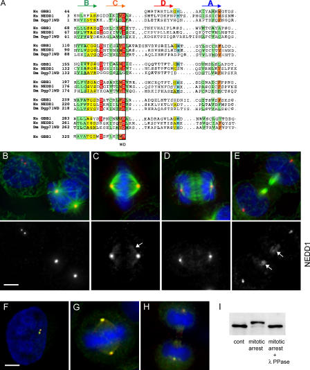
Figure 1.
NEDD1 is recruited to the mitotic centrosome and to spindle microtubules and colocalizes with γ-tubulin. (A) Alignment of the WD repeats of human NEDD1 (available from GenBank/EMBL/DDBJ under accession no. 74762597) and Dgp71WD (accession no. 28628541), together with those of human transducin β chain 1 (GBB; accession no. 51317302). The positions of predicted β strands (A, B, C, and D) are indicated above the alignment. Green and orange boxes indicate conservation of hydrophobic and aromatic residues, respectively. Additional sequence hallmarks found in WD repeats are indicated in yellow, red, and blue. (B–E) HeLa cells immunostained for NEDD1 (red), α-tubulin (green), and DNA (blue). NEDD1 is enriched at the mitotic centrosome and is found along spindle and midbody microtubules (arrows). (B) Interphase and prometaphase cells; (C) metaphase cell; (D) anaphase cells; (E) telophase cell. Bar, 5 μm. (F–H) Cells stained for NEDD1 (red), γ-tubulin (green), and DNA (blue). NEDD1 colocalizes with γ-tubulin throughout cell cycle, at interphase (F), metaphase (G), and telophase (H). Bar,5 μm. (I) HeLa cell lysates immunoblotted for NEDD1: untreated cells (cont), after overnight incubation in 500 nM taxol (mitotic arrest), or lysate of cells in mitotic arrest treated with λ-phosphatase (400 U for 30 min at 30°C).

