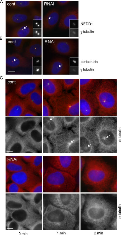
Figure 4.
Silencing of NEDD1 in interphase cells affects centrosomal localization of γ-tubulin and microtubule nucleation. (A and B) HeLa cells treated with control siRNA (cont) or NEDD1 siRNA (RNAi) and stained for NEDD1 or pericentrin (green), γ-tubulin (red), and DNA (blue). Centrosomes appear in yellow in control cells because NEDD1 or pericentrin colocalize with γ-tubulin. Insets show enlarged views of the centrosomes indicated by arrows. (C) Microtubule regrowth assay in U2OS cells treated with control siRNA or in cells treated with siRNA against NEDD1. Cells were cold treated and reheated for 0, 1, or 2 min at 37°C before staining for NEDD1 (green), α-tubulin (red), and DNA (blue). Arrows point to microtubule asters nucleated in control cells; in RNAi-treated cells, asters are barely visible. Bars, 10 μm.

