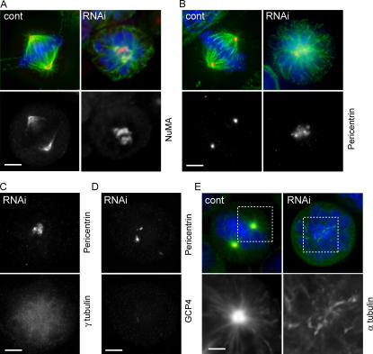
Figure 6.
Dispersion of the microtubule organization centers in NEDD1-depleted mitotic cells. (A and B) HeLa cells stained for α-tubulin (green) and DNA (blue), NuMA (A, red) as a marker of microtubule minus ends, and pericentrin (B, red) as a marker of the pericentriolar material. NEDD1-depleted cells (RNAi) show poorly separated and disorganized spindle poles. (C and D) NEDD1-depleted cells stained for pericentrin and the γTuRC proteins γ-tubulin (C) and GCP4 (D). (E) Microtubule regrowth assay in mitotic cells. Cells were cold treated and reheated for 30 s at 37°C before staining for NEDD1 (green), α-tubulin (red), and DNA (blue). Magnified areas show microtubules nucleated from the centrosome in control cells and from dispersed sites in silenced cells. Bars, 5 μm.

