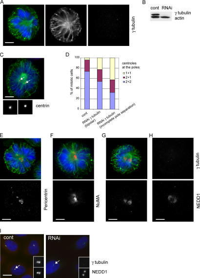
Figure 8.
Silencing of γ-tubulin inhibits centriole duplication and causes spindle defects similar to those of NEDD1 depletion. (A) Mitotic HeLa cell depleted of γ-tubulin and stained for α-tubulin (green), γ-tubulin (red), and DNA (blue). Bar, 5 μm. (B) Immunoblot of lysates (40 μg) of cells treated with control siRNA (cont) or γ-tubulin siRNA (RNAi) and probed with antibodies against γ-tubulin and actin. (C) A depleted cell stained for centrin (red), α-tubulin (green), and DNA (blue). Insets show magnified views of centrin staining of left and right spindle poles, respectively. Arrows indicate positions of the centrosomes. Bar, 5 μm. (D) Quantification of centrin-stained centrioles per pole (mean of two experiments; 232 control RNA-treated cells, 35 γ-tubulin–depleted cells with fully separated poles, and 180 depleted cells with incompletely separated poles were scored). (E–G) Cells depleted of γ-tubulin and stained in green for α-tubulin and in red for pericentrin (E), NuMA (F), and NEDD1 (G). (H) Cell depleted of γ-tubulin and stained for γ-tubulin and NEDD1. Mitotic cells depleted of γ-tubulin show disorganized spindle poles and diffuse staining of NEDD1. (I) Control and γ-tubulin–depleted interphase cells (RNAi) stained for γ-tubulin (red), NEDD1 (green), and DNA (blue). NEDD1 is concentrated at the centrosome (arrows and bottom insets), even in the absence of γ-tubulin (top insets).

