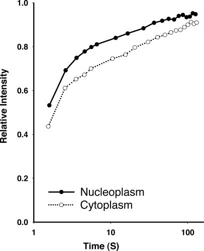
Figure 5.
Recovery of GFP-actin in the cytoplasmic and nuclear compartments. HeLa cells stably expressing GFP–β-actin were analyzed by FRAP. FRAP data was collected from cells where either the cytoplasm or the nucleoplasm was photobleached. The mean recovery profile is plotted versus recovery time. Time is plotted on a log scale to better illustrate both the rapid and the slow populations of molecules.

