Figure 1.
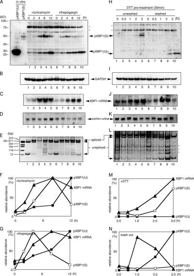
pXBP1(U) expression is induced during the recovery phase of ER stress. (A and B) Accumulation of pXBP1(U) and pXBP1(S) upon ER stress. HeLa cells were treated with 2 μg/ml tunicamycin (Tm) or 300 nM thapsigargin (Tg) for the indicated period, and cell lysates were subjected to immunoblotting with anti–XBP1-A (A) and anti-GAPDH antisera (B). In vitro–translated pXBP1(U) and pXBP1(S) were included for comparison. (C and D) Accumulation of XBP1 mRNA upon ER stress. Total RNA from cells treated as described in A was subjected to Northern blotting with XBP1 (C) and GAPDH (D) cDNA probes. (E) Splicing of XBP1 pre-mRNA during ER stress. RT-PCR was performed using total RNA extracted from cells treated as described in A as a template and the PCR primers shown in Fig. 2 A to discriminate spliced mature XBP1 mRNA from unspliced pre-mRNA. The amplified cDNA was digested with PstI, which specifically cleaves cDNA of XBP1 pre-mRNA, and separated on polyacrylamide gel. (F and G) Quantified data for A–D. pXBP1(U) (closed box), pXBP1(S) (open box), and XBP1 mRNA (closed triangle) are shown. (H and I) Accumulation of pXBP1(U) in the recovery phase. HeLa cells treated with 1 mM DTT for 30 min were washed with PBS as indicated, and lysates were prepared and analyzed by immunoblotting with anti-XBP1-A (H) antiserum that detects both pXBP1(U) and pXBP1(S) and anti-GAPDH (I) antiserum. *, nonspecific bands. (J and K) Accumulation of XBP1 mRNA during the recovery phase. Total RNA isolated from cells treated as in H was subjected to Northern blotting with XBP1 (J) and GAPDH (K) cDNA probes. (L) Splicing of XBP1 pre-mRNA in the recovery phase. RT-PCR was performed with total RNA as in E. (M and N) Quantified data for H–K. pXBP1(U) (closed box), pXBP1(S) (open box), and XBP1 mRNA (closed triangle) are shown.

