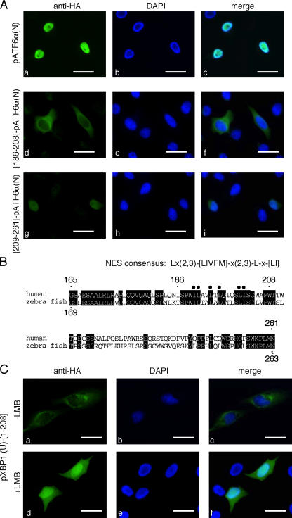
Figure 5.
pXBP1(U) contains a conventional NES. (A) NES of pXBP1(U) is functional when fused with a heterologous protein. Localization of pATF6α(N) fused with the indicated regions of pXBP1(U) was analyzed as in Fig. 3 A. (B) Comparison of amino acid sequences between human pXBP1(U)-[165–261] and the corresponding region of zebrafish pXBP1(U). Identical residues are shaded, and conserved hydrophobic residues are marked. (C) Nuclear export of pXBP1(U) is inhibited by LMB. HeLa cells transiently transfected with pCMV-HA-pXBP1(U)-[1–208] were treated with 10 nM LMB for 2 h and analyzed by immunofluorescence microscopy as in A. Bars, 10 μm.

