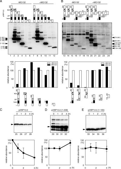
Figure 6.
Identification of the region responsible for rapid degradation. Cells were transfected with plasmid expressing the indicated deletion mutants of HA-tagged pXBP1(U) and treated with 10 μM MG132 for 2 h as indicated. Cell lysate was analyzed by Western blotting using anti–XBP1-A antiserum (A) and anti-HA serum (B). (bottom) Data quantified by an image analyzer. (C–E) Stability of pXBP1(U) and its mutants. Cells expressing pXBP1(U) (left), pXBP1(U)-[1–208] (middle), and pXBP1(U)-[1–185] (right) were treated with 40 μM cycloheximide for the indicated period, and cell lysates were subjected to immunoblotting with anti–XBP1-A antiserum. (bottom) Averages from two independent experiments are presented with standard deviation values.

