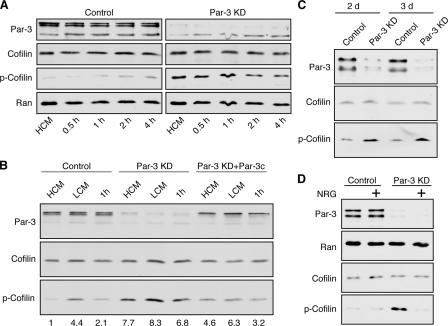
Figure 1.
Loss of Par-3 leads to elevated levels of phospho-cofilin. (A) MDCK cells were transfected with control or pSUPER–Par-3 (Par-3 KD) to suppress Par-3 expression followed by calcium switch. HCM, high calcium medium. Total cell lysates were analyzed by immunoblotting. (B) Ectopic Par-3c reduces phospho-cofilin levels in Par-3–depleted cells. A construct encoding human Par-3c was cotransfected with pSUPER–Par-3 into MDCK cells followed by calcium switch and Western blot analysis of total cell lysates. Numbers indicate relative levels of phospho-cofilin normalized against the total cofilin level. LCM, low calcium medium. (C) Depletion of endogenous Par-3 in HeLa cells increases phospho-cofilin levels. HeLa cells were transfected with human Par-3–specific (Par-3 KD) or control siRNAs. Cell lysates were prepared 48 and 72 h later and blotted with indicated antibodies. (D) NRG treatment abolishes the increase in phospho-cofilin in Par-3–depleted MCF-7 cells. 1 d after transfection with Par-3–specific or control siRNAs, MCF-7 cells were serum starved overnight followed by 45 min of treatment with 50 ng/ml NRG in serum-free medium before cells were lysed.

