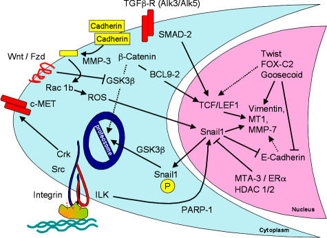
Figure 1.
Signaling events during EMT. The major signaling events that were reported in the meeting are summarized. Cleavage of E-cadherin (yellow) by MMP-3 resulted in activation of Snail1 through ROS. Snail1 localization to the nucleus is controlled by phosphorylation of a nuclear export motif and a proteosomal degradation motif, which are each phosphorylatable by GSK-3β. An ILK-responsive element in the Snail1 promoter binds PARP-1. Snail1 expression is inhibited by the MTA3–NuRD chromosomal rearrangement complex, acting downstream of the activated estrogen receptor. Repression of E-cadherin by Snail1, Twist, or other repressors leads indirectly to expression of vimentin and other mesenchymal gene products, partly because of β-catenin/Tcf–Lef1 activation. FOX-C2, as well as SIP1, can also directly activate mesenchymal gene expression. Translocation of β-catenin to the nucleus requires BCL9-2, which itself can induce EMT. Abundance of β-catenin is regulated by phosphorylation-dependent proteosomal degradation, unless GSK-3β is silenced through Wnt signaling. TGF-β is known to activate this canonical Wnt pathway, but TGF-β also directly activates the Tcf–Lef1 transcription complex through tyrosine phosphorylation of SMAD-2. The c-Met receptor tyrosine kinase, through the Crk adaptor, also stimulates EMT.

