Figure 1.
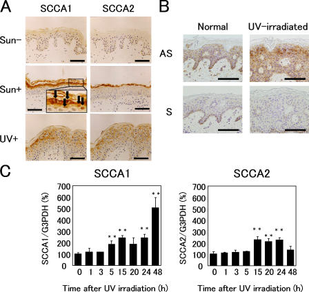
SCCA is up-regulated by UV irradiation in vivo and in vitro. (A) Immunostaining for SCCA1 and SCCA2 in sun-protected buttock skin, sun-exposed (cheek), and UV-irradiated buttock skin (two minimal erythema doses of radiation; biopsy taken after 48 h). The inset shows nuclear localization of SCCA1 at high magnification. Arrows indicate heavy SCCA1 staining in the nuclei. (B) In situ mRNA hybridization of SCCA1. The sense probe did not show any positive reaction. The dark brown color seen in basal cells is caused by melanin. (C) Quantitative real-time PCR analysis of SCCA1 and SCCA2 mRNA levels in cultured human keratinocytes. Values given are SCCA1 or SCCA2 mRNA levels normalized to the amount of G3PDH. Error bars represent the mean of five wells ± SD. **, P < 0.01. Bars, 100 μm.

