Figure 2.
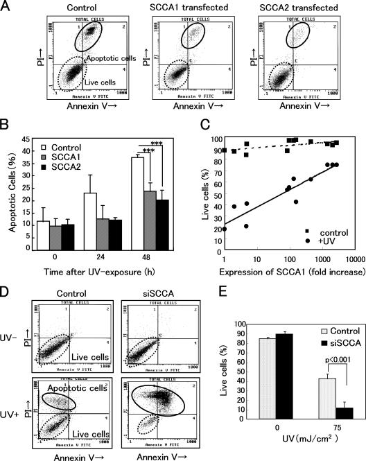
Overexpression or down-regulation of SCCAs significantly affected UV-induced apoptosis. (A) FACS analyses of apoptotic cells using SCCA1- or SCCA2-transfected 3T3/J2 cells, 48 h after UV irradiation (30 mJ/cm2). Cells were stained with FITC-conjugated Annexin V and propidium iodide. (B) Analyses of five experiments are summarized. (C) 12 clones whose expression levels of SCCA1 mRNA distributed from 1 to 2,772-fold were established. Using these clones, the effects of UV irradiation were examined. Cells were harvested 48 h after UV irradiation (50 mJ/cm2) and FACS analyses were performed. The antiapoptotic activity correlated with SCCA1 expression. r = 0.734. (D) Using pSilencer vector, an siRNA construct targeted to a homologous sequence of SCCAs was stably transfected into HaCaT keratinocytes. Typical FACS analyses of nonirradiated (UV−) and UV-irradiated (UV+) siSCCA/HaCaT cells were shown. Apoptotic cells were analyzed 48 h after UV irradiation (75 mJ/cm2). (E) Statistical analyses of five experiments. Error bars represent the mean of five wells ± SD.

