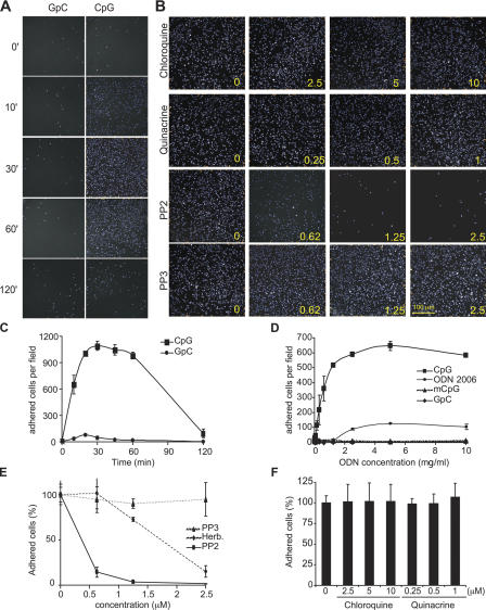
Figure 3.
CpG-induced cell adhesion is insensitive to chloroquine but requires SFK activity. (A) Cells were stimulated with 1 μg/ml of a control GpC-ODN or with CpG–ODN 2216 for the indicated times (min). After fixation, cells were visualized as described in Materials and methods. (B) THP-1 cells were preincubated for 10 min with chloroquine, quinacrine, PP2, and PP3. The number at the lower right corner of each panel indicates the concentration for each inhibitor used (μM). Cells were then stimulated for 30 min with CpG, and adhesion was measured. Representative fields are presented. (C) THP-1 cells were stimulated as indicated in A, and images captured from 36 different fields from at least four different wells were quantified to obtain cell number per field and plotted with the SD. (D) THP-1 cells were stimulated for 20 min with increasing concentrations of CpG, control GpC, the B class oligo 2006 (ODN 2006), and murine CpG (mCpG), and adhesion was measured. (E) Adhesion was measured on THP-1 cells that were pretreated for 10 min with increasing concentrations of PP2, PP3, and herbimycin A and then stimulated with CpG for 30 min. Data are represented as percentage of adhered cells versus vehicle-pretreated cells. (F) THP-1 cells were preincubated for 10 min with increasing concentrations of chloroquine or quinacrine. Cells were then stimulated for 30 min with CpG, and the number of adhered cells from at least 36 different fields was quantified. Error bars indicate SD.

