Figure 7.
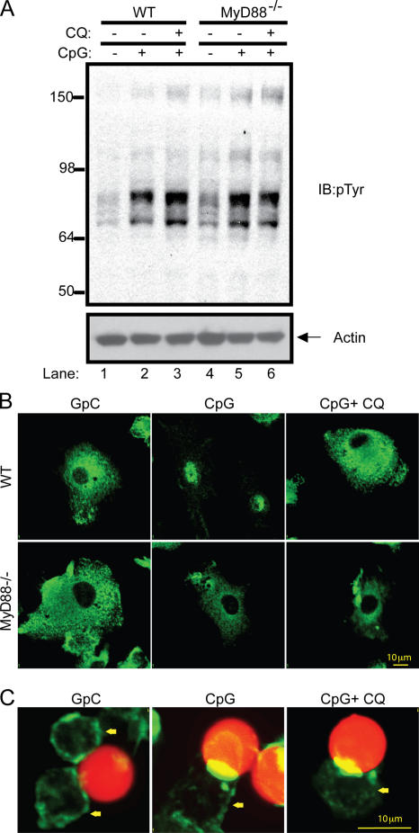
CpG activates SFKs at the plasma membrane and upstream of the MyD88 activation. (A) Thioglycollate-elicited peritoneal macrophages were obtained from wild-type and MyD88−/− mice and cultured for 24 h in medium containing 0.5% FBS. Cells were pretreated with 10 μM chloroquine (CQ) or vehicle for 15 min and stimulated with CpG–ODN 1585 for 5 min. Tyrosine phosphorylation after CpG treatment was visualized by immunoblotting with an anti-pTyr antibody. (B) Murine bone marrow–derived DCs from wild-type and MyD88−/− mice were pretreated with 10 μM chloroquine or vehicle for 15 min, and after CpG stimulation, cells were stained with an antibody to the p65 subunit of NF-κB. (C) CpG- or GpC-coated 10-μm red fluorescent polystyrene beads were added to RAW cells for 20 min. Cells were then fixed and stained with FITC-phalloidin. The yellow arrow indicates the location of cells.

