Figure 1.
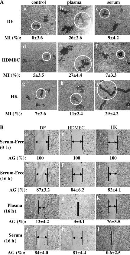
Plasma and serum oppositely affect dermal and epidermal cell migration. HKs, DFs, and HDMECs were serum starved overnight and subjected to colloidal gold migration assays (A) and in vitro wound-healing assays (B). (A) Representative images of the colloidal gold migration assays under the following three conditions: (1) on a collagen matrix in the absence of any soluble GFs (a, d, and g); (2) on a collagen matrix in the presence of an optimal concentration (10% vol/vol) of human plasma (b, e, and h); and (3) on collagen matrix in the presence of an optimal concentration (10% vol/vol) of human serum (c, f, and l). The computer-assisted quantitative analyses of the migration tracks are shown as migration indices (MI). These experiments were repeated five times, and closely similar results were obtained. Average size migration tracks are marked with circles. (B) In vitro wound-healing assay was performed under the same conditions. The wound closures were photographed, and the average gaps (AGs) were quantitated as described previously (Li et al., 2004b). AGs are indicated by lines and arrows; some gaps were too close to insert an arrow. Similar results were reproduced in three independent experiments.

