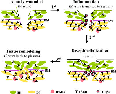
Figure 8.
A schematic representation of how the plasma→serum→plasma transitions coordinate the orderly skin cell migration during wound healing. Three major types of skin cells—HKs, DFs, and HDMECs—are shown here, as indicated by different colors. The dermal cells express higher levels of TβRII (symbolized by Y) than the epidermal cells. Therefore, the dermal cells are sensitive to the anti-promotility effect of TGFβ3 (red stars), whose concentration increases after the transition from plasma to serum in the wound bed. Contributions by other cell types and matrix components to skin wound healing are omitted for the sake of simplicity but not reality. The relative numbers and proportions of the various types of cells do not quantitatively reflect those in real human skin. BM, basement membrane.

