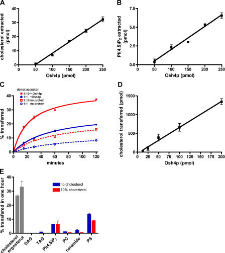
Figure 3.
Extraction and transfer of sterol from liposomes by purified Osh4p. (A) 50 μL of 1 mM liposomes containing DOPC and 14C-cholesterol (99:1 mol%) in TS were mixed with the indicated amount of Osh4p at 30°C for 1 h. The amount of 14C-cholesterol extracted was determined as described in Materials and methods. (B) Liposomes containing DOPC and 3H-PI(4,5)P2 (99:1 mol%) in TS were incubated with the indicated amount of Osh4p for 1 h at 30°C. The amount of 3H-PI(4,5)P2 extracted was determined. (C) Donor vesicles (DOPC/egg PE/lactosyl-PE/14C-cholesterol/PI(4,5)P2; 59.5:20:10:10:0.5 mol%) were mixed with acceptor vesicles (DOPC/PE; 80:20 mol%) at a molar ratio of 1:10 or 1:1, mixed with 40 picomols (pmol) of Osh4p, and incubated at 30°C. At various times, samples were place on ice and the amount of 14C-cholesterol transferred to the donor was determined. The amount of 14C-cholesterol transferred in the absence of protein is also shown. (D) Donor vesicles were prepared as described in C and mixed with acceptor membranes at a molar ratio of 1:10. Different amounts of Osh4p were added and the reactions were incubated at 30°C for 15 min. The amount of radiolabeled sterol transferred to acceptor membranes minus the amount transferred in the absence of protein was determined. (E) Donor membranes were prepared as in C with either 10% 14C-cholesterol or 10% 3H-ergosterol (gray bars) or with either 1.0% 14C-DAG, 0.01% 3H-TAG, 0.5% 3H-PI(4,5)P2, 0.008% 3H-PC, 1.0% 14C-ceramide, or 10% 3H-PS and either no (blue bars) or 10% (red bars) cholesterol. These vesicles were mixed with acceptor vesicles at molar ratio of 1:10 and incubated with 40 pmol Osh4p at 30°C for 1 h. The amount of radiolabeled lipid transferred to acceptor membranes was calculated by subtracting the amount of transfer in control reactions incubated at 4°C. In all graphs, the values are the mean of three determinations. Error bars indicate the SEM. n = 3.

