Figure 4.
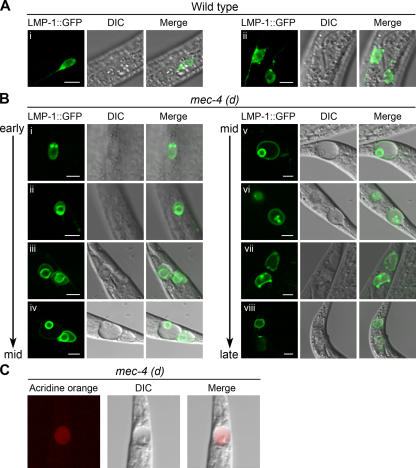
Lysosomal morphology and distribution during neurodegeneration. (A) Confocal images of wild-type touch receptor neurons expressing a pmec-17 LMP-1∷GFP transgene. LMP-1∷GFP expression, differential interference contrast (DIC), and merged images are shown. Healthy neurons show a scattered and punctate pattern of lysosomal distribution. (i) Wild-type PVM touch receptor neuron. (ii) Wild-type ALM (left and right) touch receptor neurons. (B) Confocal images of PLM touch receptor neurons of mec-4(d) animals expressing a pmec-17LMP-1∷GFP transgene. During the early to middle (mid) stages of degeneration, lysosomes enlarge and appear to coalesce around a swollen nucleus (i–iv). Later on, the nucleus migrates to the periphery of the cell and condenses (iv–vi). At the late stage, no lysosome structure is evident and the vacuolated cell becomes diffusely fluorescent (vii and viii). (C) Acridine orange staining of a middle to late degenerating PLM touch receptor neuron. Acridine orange, DIC, and merged images are shown. Bars, 5 μm.

