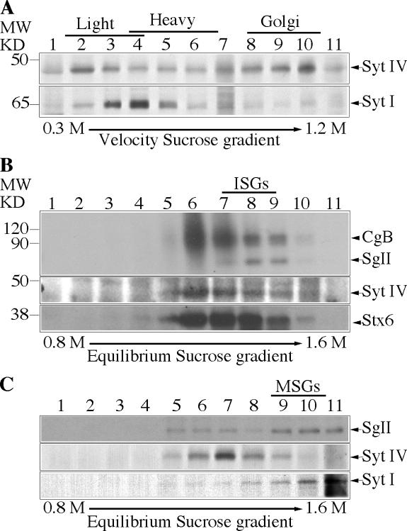
Figure 1.
Syt IV is found on the Golgi and ISGs in PC12 cells. (A) Fractions from a 0.3–1.2 M continuous velocity sucrose gradient loaded with a PNS from PC12 cells were collected and analyzed with anti–Syt IV (top) and Syt I (bottom) antibodies. Alternatively, PC12 cells were labeled with [35S]sulfate for 5 min and chased for 15 min (B) or labeled for 1 h and chased O/N (C) to label ISGs or MSGs, respectively, and then subjected to velocity centrifugation. (B and C) Fractions 1–4 (B) or 4–6 (C) from the velocity gradients were further separated on a 0.8–1.6 M discontinuous equilibrium sucrose gradient and analyzed. (B and C, top) Autoradiogram showing [35S]sulfate-labeled CgB (B) and SgII (B and C). (B and C, bottom) Western blot analysis using anti–Syt IV and anti-Stx6 antibodies (B) or anti–Syt IV and anti–Syt I antibodies (C). In B, the diffuse band between 120 and 90 kD is a heparan sulfate proteoglycan (Tooze and Huttner, 1990).

