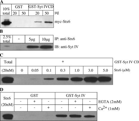
Figure 4.
Syt IV interacts with Stx6. (A) Cell lysates from HEK cells transfected with myc-Stx6 were incubated with GST-Syt IV CD or GST bound to glutathione beads. (B) PC12 cells were lysed in TNTE buffer and incubated with 5 or 10 μg of anti-Stx6 antibody overnight at 4°C, and subsequently bound to protein G beads. Immunoprecipitates and 2.5% of the input were analyzed by immunoblotting with anti–Syt IV antibody. (C) Direct binding of Stx6 to Syt IV in vitro. 3 μg of GST-Syt IV CD bound to glutathione beads was incubated with the indicated amounts of the Stx6 CD in binding buffer overnight, and bound protein was analyzed by immunoblotting with anti-Stx6 antibody 3D10. (D) The Syt IV–Stx6 interaction in vitro is independent of Ca2+. Binding assays were performed as in C, using 1 μM Stx6 CD, supplemented with either 1 mM CaCl2 or 2 mM EGTA.

