Figure 6.
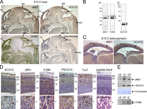
Expression of JNK1 and SCG10 overlap spatially in developing mouse brain. If SCG10 is a physiological target for JNK, it should localize regionally with active JNK. (A) To test this, sagittal sections of embryonic day (E) 15.5 mouse embryos were immunostained with antibodies recognizing JNK1, SCG10, and GFAP or with preimmune serum for SCG10. Immunoreactivity was detected with 3,3′-diaminobenzedine tetrahydrochloride (brown) and counterstained with hematoxylin (blue). Regions displaying prominent expression of JNK1 and SCG10 are indicated. TE, telencephalon; OE, olfactory epithelium; MB, roof of the midbrain; IC, inferior colliculus; MO, medulla oblongata. Bar, 500 μm. (B) Specificity of the antibodies toward forebrain extract. (C) The telencephalon is divided into the marginal zone (MZ), cortical plate (CP), intermediate zone (IZ), and ventricular zone (VZ) as indicated. Bar, 200 μm. (D) Closer inspection of the telencephalon confirms that SCG10, JNK1, PJNK, PSCG10, and class III β-tubulin (TuJ1) immunoreactivity localizes to the intermediate zone, cortical plate, and marginal zone (arrows) and is predominantly cytosolic (bottom; arrows). Preincubation of α-PSCG10 with phospho-S73 antigenic peptide blocks immunoreactivity. Bar, 50 μm. (E) To verify that the immunoreactivity observed corresponded to the antigens tested, multiple sections of the cortical plate, intermediate zone, and ventricular zone were microdissected and levels of JNK1, PJNK, and SCG10 examined by immunoblotting. Protein loading is indicated by Coomassie staining of histones.

