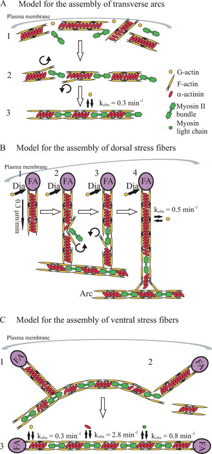
Figure 8.
Model for the assembly of transverse arcs, dorsal stress fibers, and ventral stress fibers. (A) Transverse arc. (1) Short actin bundles cross-linked by α-actinin are generated at the plasma membrane through nucleation by the Arp2/3 complex. (2) Actin bundles associate endwise with myosin II bundles close to the plasma membrane. (3) During centripetal flow, the α-actinin and myosin II bands are equalized in width, the entire bundle is straightened, and the width of bands is decreased, apparently as the result of a contraction of the transverse arc. (B) Dorsal stress fiber. (1) After formation of a focal adhesion (FA), short unipolar actin filaments are polymerized by mDia1/DRF1-dependent mechanism (Dia) from the focal adhesion at the rate of 0.3 μm/min. Polymerized actin filaments are simultaneously cross-linked by α-actinin. (2–4) The proximal end of a dorsal stress fiber is connected to the side of a transverse arc. When dorsal stress fiber has reached the length of several micrometers, myosin II can be occasionally incorporated into α-actinin cross-linked bundle, leading to a simultaneous displacement of α-actinin. (C) Ventral stress fiber. (1) Preassembled dorsal stress fibers and arcs interact with each other. (2) The transverse arc region that is not located between the two dorsal stress fibers is disconnected from the structure. (3) The transverse arc aligns between the two dorsal stress fibers, contracts, and subsequently forms a ventral stress fiber that is anchored to focal adhesions at both ends. The dynamics of actin, α-actinin, and MLC association to stress fibers are indicated in the figure. Note that α-actinin associates with stress fibers in a highly dynamic manner. This dynamic filament cross-linking may be essential for myosin incorporation into dorsal stress fibers as well as for contractility of mature stress fibers.

