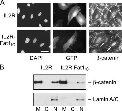
Figure 8.
Effect of Fat1IC overexpression on β-catenin nuclear localization in VSMCs. (A) Immunofluorescence analysis of β-catenin subcellular localization in IL2R-GFP-RV (top) and IL2R-Fat1IC-GFP-RV (bottom) transduced RASMCs. Cells were treated with LiCl (20 mmol/L) for 6 h, and then stained with anti-β-catenin antibody and DAPI. Transduced cells were identified by coexpressed GFP. Arrows indicate nuclear β-catenin signal of untransduced and transduced cells within each panel (see text). Bar, 10 μm. (B) Western analysis of β-catenin in membrane (M), cytoplasmic (C), and nuclear (N) fractions extracted from IL2R-GFP-RV and IL2R-Fat1IC-GFP-RV transduced A7r5 cells treated with LiCl, as above. The blot was probed for lamin A/C to assess fractionation and loading.

