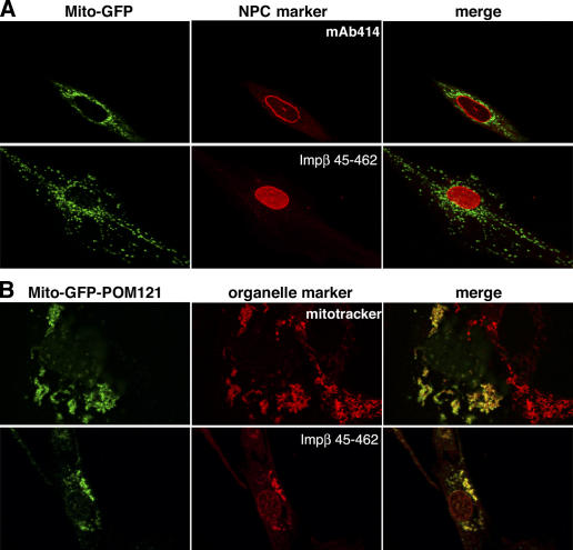
Figure 1.
Ectopic expression of POM121 at mitochondria. (A) EGFP was fused behind residues 1–70 of TOM20, expressed in HeLa cells, and thereby anchored to the outer mitochondrial membrane (Mito-GFP). (top) Colocalization of the fusion protein with the NPC marker mAb414. (bottom) Staining of NPCs with the Alexa 568–labeled Impβ45–462 after digitonin permeabilization. Signals for mitochondria and NPCs do not overlap. (B) The POM12173–797 fragment, lacking its natural membrane anchor and the FG repeat domain, was fused behind the Mito-GFP module (Mito-GFP-POM121) and expressed in HeLa cells. (top) Colocalization of the Mito-GFP-POM121 signal with mitotracker-stained mitochondria (images depict a transfected and a nontransfected cell). (bottom) Bright staining with Impβ45–462 indicates recruitment of FG or GLFG repeat Nups to the ectopic POM121 fragment at mitochondria. Clustering of mitochondria is a side effect of this ectopic expression. The weak Mito-GFP-POM121 signal at the NE reflects the fact that targeting of the fusion protein to mitochondria is in competition with incorporation into bona fide NPCs.

