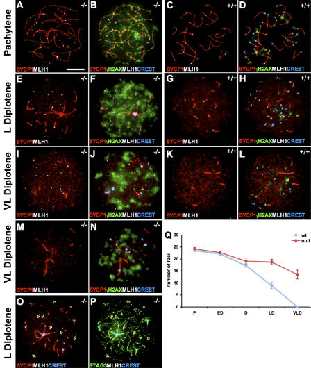
Figure 5.
Absence of SYCP3 delays the removal of MLH1 during meiosis. The distribution of MLH1 foci (white) and association with synapsed regions (labeled by SYCP1; red) at different stages of meiosis are shown for Sycp3 −/− oocytes (C, G, K, and M), whereas wild-type oocytes are shown as a control (A, E, and I). γH2AX (green) and centromeres visualized by CREST (blue) were added to these pictures for Sycp3 −/− oocytes (D, H, L, and N) and wild-type oocytes (B, F, and J). A subset of the Sycp3 −/− oocytes displays fewer foci than the rest (a very late diplotene stage oocytes is shown as an example [M and N]). A majority of the MLH1 foci in late diplotene Sycp3 −/− oocytes was not associated with SYCP1 staining (O, arrows), but was associated with the asynapsed axial cores as defined by the cohesin complex protein STAG3 (P, arrows). (Q) The number of MLH1 foci was presented as the mean ± SEM of analyzed oocytes from five meiotic stages (n a, counted wild-type oocytes; n b, counted Sycp3 −/− oocytes). The number of MLH1 foci shows no difference between wild-type and Sycp3 −/− oocytes at the pachytene (middle to late; A and C; n a = 50; n b = 35) and early (n a = 36; n b = 41) to middle diplotene (n a = 24; n b = 29). An increased number of MLH1 foci relative to wild-type is seen in late (G) and very late diplotene (K) Sycp3 −/− oocytes, (LD, n a = 20 and n b = 42; VLD, n a = 20 and n b = 24). Bars, 10 μm.

