Figure 5.
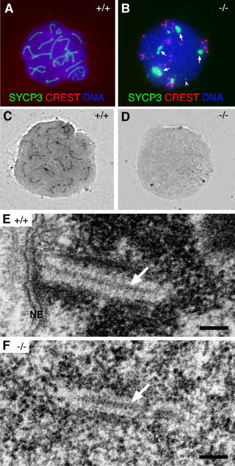
Analysis of SC formation in Sycp2−/− spermatocytes. Surface-spread nuclei were stained with anti-SYCP3 antibodies, CREST, and DAPI, or they were stained with silver nitrate. (A) SYCP3 labels SCs in wild-type spermatocytes. (B) SYCP3 fails to incorporate into SCs in Sycp2 −/− spermatocytes. SYCP3 forms several large protein aggregates (arrows). In addition, SYCP3 also forms many small nuclear foci (arrowhead). (C) SCs in wild-type spermatocytes are readily stained with silver nitrate. (D) No silver-stained SC structures are visible by light microscopy in Sycp2 −/− spermatocytes. (E) EM analysis of a wild-type spermatocyte. A tripartite structure of SC is formed by one CE (arrow) that connects with two electron-dense LEs. This SC is attached to the nuclear envelope (NE). Note the condensed chromatin around the SC. (F) EM analysis of a Sycp2 −/− spermatocyte. TFs form a CE-like structure (arrow). Two rows of chromatin grains align in parallel with the CE-like structure to form a SC-like structure. Bars, 0.2 μm.

