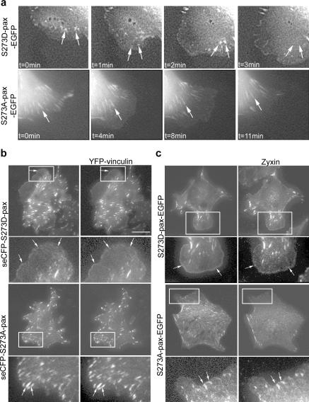
Figure 3.
S273-paxillin phosphorylation induces the formation of small, dynamic adhesions in protrusions. (a, top) Time-lapse imaging of CHO-K1 cells showed S273D-paxillin-GFP localization in small adhesions that appeared and disappeared (turned over) in 1–2 frames (arrows) near the leading edge. (bottom) On the other hand, S273A-paxillin-GFP localized into large adhesions (arrow) near the base of the protrusion that either tend to slide or disassemble slowly (Table I). (b, top) seCFP-S273D-paxillin and YFP-vinculin exhibit colocalization in small adhesions within protrusions (arrows) of CHO-K1 cells, imaged by TIRF. Enlargements of the boxed regions are at the bottom of each panel. Rates of paxillin and vinculin disassembly in these adhesions were accelerated similarly (Table I), indicating that fast turnover is a property of the entire adhesion. (bottom) In contrast, CHO-K1 cells coexpressing seCFP-S273A-paxillin and YFP-vinculin exhibited larger adhesions (arrows) with similar but slower adhesion disassembly rates (Table I). Bar, 20 μm. (c) S273A- and S273D-paxillin-GFP colocalized with zyxin in small adhesions in protrusive parts of CHO-K1 cells and in larger adhesions at the protrusion base, respectively, as shown in TIRF experiments. Arrows point to the regions where paxillin and zyxin colocalize.

