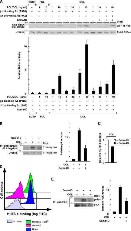
Figure 2.
Sema4D inhibits ECM-mediated activation of R-Ras and functional activation of β1 integrins. (A) PC12 cells were collected and kept in suspension (SUSP) or replated onto poly-d-lysine (PDL)– or collagen (COL)–coated (1 or 10 μg/ml) dishes with or without Sema4D in the plating media. 15 min after plating, the cells were lysed and the lysates were incubated with GST-RBD, and bound R-Ras protein and total lysates were analyzed by immunoblotting. For the indicated samples, cells were treated with 5 μg/ml of monoclonal β1 integrin blocking (P5D2) or activating (8A2) antibody before replating. Relative R-Ras activity was determined by the amount of R-Ras bound to GST-RBD normalized to the amount of R-Ras in cell lysates analyzed by NIH Image software (bottom). (B and E) PC12 cells were seeded onto noncoated or collagen-coated (10 μg/ml) dishes, with or without Sema4D in the plating media. Cells were lysed, and the lysates were immunoprecipitated with an antibody against the active β1 integrins, HUTS-4 (B), or an antibody against FAK (E) to measure the activity of β1 integrins or tyrosine phosphorylated FAK, respectively. (C) The ELISA using HUTS-4 antibody was performed to confirm the effect of Sema4D on activity of β1 integrins under a detergent-free condition. Results are the means ± SEM of three independent experiments. (D) PC12 cells were treated for 3 h at 37°C with control medium or with medium containing Sema4D or Sema4D plus 1 mM Mn2+. Cells were incubated with HUTS-4 antibody or buffer alone (−1st Ab), followed by labeling with the FITC-conjugated secondary antibody. Fluorescence intensity was determined by flow cytometry analysis. Error bars indicate SEM.

