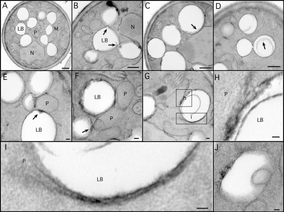
Figure 2.
Ultrastructure of lipid bodies and contacts with peroxisomes. Cells were grown for 18–21 h in oleate medium and processed for EM. (A–D) Aspects of lipid body structure are illustrated, particularly their connections to each other (A and B) and inclusions (C and D). Arrows in B illustrate valvelike structures; arrows in C and D illustrate inclusions. (E–H) Lipid bodies in contact with peroxisomes are shown, illustrating electron-dense material at the site of contact (arrows). Note a peroxisome in contact with three lipid bodies (E). A lipid body making contact with two peroxisomes, one of which has extended a tail around it, and the other has breached the periphery and is extending a process (pexopodium) into the core (G). The boxed areas are shown in higher resolution in H and I. Another putative pexopodium is shown in J. LB, lipid body; M, mitochondria; N, nucleus, P, peroxisome. Bars (A–D), 500 nm; (E–J), 50 nm.

