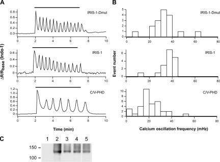
Figure 3.
Effects of exogenously expressed proteins on Ca2+ oscillation frequency in mGluR5a-expressing HeLa cells. (A) Emission ratio change of Indo-1 signals in cells stimulated with 100 μM of glutamate (horizontal bars). (B) Histograms of Ca2+ oscillation frequency in mGluR5a-expressing cells stimulated with 100 μM of glutamate. (C) Western blot analysis of cell lysates prepared from HeLa cells transfected with mGluR5a alone (lane 2), mGluR5a plus C/V-PHD (lane 3), mGluR5a plus IRIS-1 (lane 4), or mGluR5a plus IRIS-1–Dmut (lane 5). Nontransfected cells were used as a control (lane 1). An anti-mGluR5 antibody was used. Molecular mass markers are shown on the left (×10−3).

