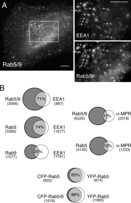
Figure 5.
Colocalization of Rab5/9 with EEA1. (A) Colocalization of CFP-Rab5/9 with the early endosome marker EEA1 in fixed HeLa cells stained with anti-EEA1 and anti-GFP antibodies. The square in A is enlarged for both markers (right); arrows are included to facilitate comparison. Bar, 10 μm. (B) Quantitation of CFP-Rab5/9, CFP-Rab5, and YFP-Rab9 colocalization. Venn diagrams are shown as follows: CFP-Rab5/9, CFP-Rab5, and YFP-Rab9 versus EEA1; CFP-Rab5/9 and CFP-Rab5 versus endocytosed anti–CI-MPR IgG; and CFP-Rab5/9 and CFP-Rab5 versus YFP-Rab5. The number of positive structures is indicated below each protein name; overlap is indicated as described in the Results section.

