Figure 1.
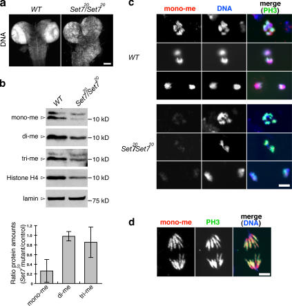
Monomethylated H4K20 is strongly reduced in PR-Set7 brains. (a) Wild-type (WT) and PR-Set7/PR-Set7 third-instar larval brains were stained with Hoechst. The two strongly staining rings (dense nuclei) observed in wild type are the optic lobes. (b) Western blots of extracts from wild-type and PR-Set7/PR-Set7 third-instar larval brains probed with anti–mono-, anti–di-, and anti–trimethylated H4K20 (mono-, di-, and tri-me), anti–histone H4, and anti-lamin antibodies. The intensity of the bands was quantified by ImageJ, and the value of mono-, di-, or trimethylated H4K20 was normalized to the values of both histone H4 and lamin (see Table S1, available at http://www.jcb.org/cgi/content/full/jcb.200607178/DC1). The graph shows the ratio of PR-Set7/PR-Set7 mutant to wild-type values. Error bars show two SDs (n = 3). (c) Neuroblasts were costained with anti–monomethylated H4K20 (mono-me; red) and anti-PH3 antibodies (green). DNA was stained with Hoechst (blue). (d) Monomethylated H4K20 (red) is distributed all along the chromosomes. Bars: (a) 100 μm; (c and d) 5 μm.

