Figure 2.
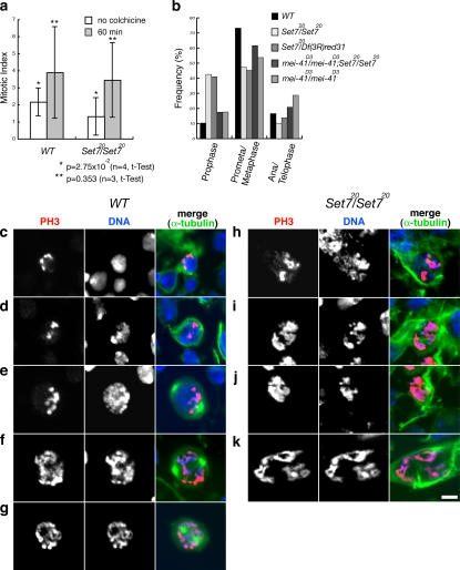
PR-Set7/PR-Set7 neuroblasts show a delay in early mitotic stages. (a) Mitotic indexes of wild type (WT) and PR-Set7/PR-Set7 before and after incubation with colchicine for 1 h. The mitotic index was determined as the number of PH3-positive cells over the total number of cells (see Table S2, available at http://www.jcb.org/cgi/content/full/jcb.200607178/DC1). (b) Quantification of mitotic parameters in wild-type (n = 272), PR-Set720/PR-Set720 (n = 275), PR-Set720/Df(3R)red31 (n = 363), mei-41D3/mei-41D3;PR-Set720/PR-Set720 (n = 365), and mei-41D3/mei-41D3 (n = 404) neuroblasts. We considered cells in prophase if they were positive for PH3 staining and showed interphase-like organization of microtubules without visible asters. Percentage was defined as the number of mitotic cells at a specific stage over total number of mitotic cells (Table S3). (c–k) Cells were costained with anti-PH3 antibody (red), Hoechst (blue), and anti–α-tubulin antibody (green). Wild-type prophase (c and d), wild-type prometaphase (e–g), and typical prophase figures observed in PR-Set7 (h–k) are shown. Bar, 5 μm.

