Figure 1.
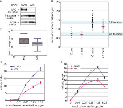
Mitotic defects in APC-deficient cells. (A) A reduction in the amount of APC protein in U2OS cells using RNAi. Lysates from U2OS cells transfected with short RNA duplexes directed against APC or with control RNA duplexes were collected 72 h after transfection. Proteins were separated by PAGE and probed for APC, β-catenin, and actin (loading control). (B) Tension between kinetochores of metaphase sister chromatids is reduced in APC-deficient U2OS cells. Interkinetochore distances in U2OS cells transfected with nontargeting (iC) or APC-targeting (iA) siRNA as in A were measured as the distances between CREST-stained kinetochores in prophase or metaphase chromosomes as indicated. Mean values are shown as red bars. (C) APC inhibition in U2OS cells decreases the time from mitotic entry to anaphase onset. U2OS cells transfected with control (iC) or APC-targeting (iA) siRNA together with H2B-RFP were presynchronized by a single thymidine block and released for 6 h before imaging their mitotic progression for 6 h. Time from initiation of mitosis to anaphase onset was measured for 14 control and 32 APC-deficient cells and is shown as box plots using a five-point summary. Pairs with a statistically significant difference (P < 0.005) in two-tailed t tests are indicated by red hashes (#). (D and E) Reducing APC causes a mitotic checkpoint defect. U2OS cells transfected for 52 h with control or APC-directed siRNA duplexes were incubated with increasing amounts of nocodazole (A) or taxol (B) for a further 20 h before staining with propidium iodide and antiphosphohistone H3 antibodies. Mitotic index was determined by flow cytometry and is shown as the percentage of live cells that were phosphohistone H3 positive and had high (∼4n) DNA content. Data are represented as the mean ± SD (error bars).

