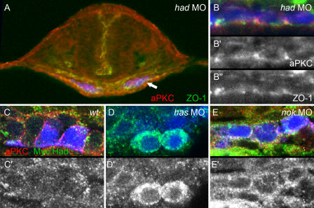
Figure 2.
Effects of had, has, and nok morphants on myocardial apical–basal polarity at the 20-somite stage. Transverse sections of heart cone stage (20-somite) embryos. GFP is false-colored in blue; aPKC, red (A–C and E) or gray (B′); Myc∷Had, green (C–E) or gray (C′–E′); ZO-1, green (A and B) or gray (B″). (A) had morphant with a section plane through the middle of the heart cone. The two bilateral wings of myocardial cells are blue. Arrow indicates the lateral portion of the myocardial field that was used for detail images within the various genetic backgrounds. (B, B′, and B″) had morphants are correctly polarized and display apical ZO-1– and aPKC-positive spots. (C and C′) In wt myocardial cells, levels of Myc∷Had/Na+,K+ ATPase are low, and a clear localization pattern is not apparent. (D and D′) has morphant myocardial cells exhibit higher levels of Myc∷Had/Na+,K+ ATPase, which is localized around the circumference of cells. (E and E′) Similarly, nok morphants display localization of Myc∷Had/Na+,K+ ATPase and aPKC around the entire myocardial circumferences, which is indicative of loss of apical–basal polarity within these cells.

