Figure 7.
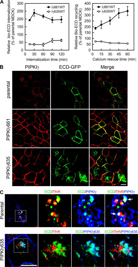
The interaction between PIPKIγ and AP1B is necessary for E-cadherin transport. (A) MDCK cells with or without PIPKIγ635 overexpression were used to analyze E-cadherin internalization and recycling by surface biotinylation as described. Immunoblot data from three independent experiments were quantified by ImageJ and plotted with SigmaPlot 8.0. Error bars are ± SEM. (B) Parental, PIPKIγ635-, or PIPKIγ661-expressing MDCK cells were transiently transfected with ECD-GFP. PIPKIγ was visualized by indirect immunofluorescence and ECD-GFP using direct fluorescence microscopy. (C, top) Parental MDCK cells were incubated at 20°C for 2 h, treated with 2 mM EGTA for 30 min, and then fixed and triple- labeled using monoclonal anti-CD71, FITC-conjugated anti–E-cadherin, and rabbit anti-PIPKIγ antibodies. (bottom) MDCK cells with the expression of HA-PIPKIγ635 induced for 72 h were maintained at 20°C for 2 h. The cells were then fixed and stained using monoclonal anti-CD71, FITC-conjugated anti–E-cadherin, and rabbit anti-HA antibodies. Arrows indicate the colocalization of ECD, PIPKIγ, and the recycling endosome. Bars, 10 μm.

