Figure 2.
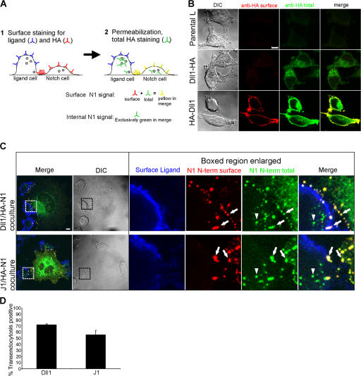
Transendocytosed Notch1 structures are internal and disconnected from the plasma membrane. (A) Staining protocol to distinguish surface and internal N1 (see Materials and methods for details). (B) Validation of protocol in A. L cells expressing Dll1 with HA tags on either the intracellular (Dll1-HA) or extracellular (HA-Dll1) domain were fixed and stained for surface HA with mouse HA antibody (262K) and anti–mouse Alexa Fluor 568 (red). After permeabilization, cells were stained for total HA (surface and intracellular) with an HA antibody (16B12) conjugated to Alexa Fluor 488 (green) and imaged by confocal and DIC microscopy. (C) Cocultures of HA-N1 cells with Dll1 or J1 cells were fixed and stained with rabbit anti-ECD antibodies to Dll1 or J1 and anti–rabbit Alexa Fluor 633 antibodies (blue) to label the surface of the ligand cell, followed by staining for surface N1 N terminus with a mouse HA antibody (262K) and anti–mouse Alexa Fluor 568 (red). After permeabilization, cells were stained for total N1 N terminus (surface and intracellular) with an HA antibody (16B12) conjugated to Alexa Fluor 488 (green), and imaged by confocal and DIC microscopy. Arrows indicate N1 N terminus on both the Notch cell surface (yellow) and the ligand cell surface (white). Arrowheads indicate internal N1 N terminus detected within ligand cells (green). Boxes denote enlarged regions. (D) Transendocytosis was quantified by examining Dll1 or J1 cells for N1 N terminus detected exclusively after permeabilization (see Materials and methods). Error bars represent the SEM. Images from each experiment were uniformly adjusted using the levels function in Photoshop. Bars, 5 μm.

