Figure 2.
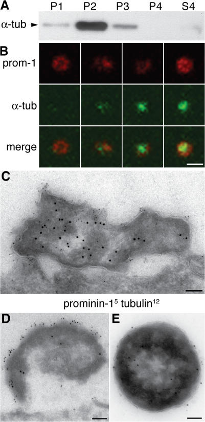
Prom1 particles contain α-tubulin. (A) Neural tube fluid of E11.5 mouse embryos was subjected to differential centrifugation (Marzesco et al., 2005) followed by immunoblot analysis of the fractions for α-tubulin. P1, 300-g pellet; P2, 1,200-g pellet; P3, 10,000-g pellet; P4, 100,000-g pellet; S4, 100,000-g supernatant. (B) Transverse cryosection of E10.5 mouse telencephalon double immunostained for prom1 (red) and α-tubulin (green) and analyzed by confocal microscopy; four examples of prom1 particles in the lumen of the telencephalic ventricle are shown (z-stack projection). (C and D) Transverse ultrathin cryosections of the apical region of mouse E10.5 telencephalic NE cells immunogold labeled for prom1 (5 nm) and α-tubulin (12 nm) showing two doubly immunoreactive particles at the lumenal surface. (E) Negative staining of a particle from the P2 pellet after immunogold labeling for prom1 (5 nm) and acetylated tubulin (12 nm). Bars: (B) 1 μm; (C–E) 100 nm.

