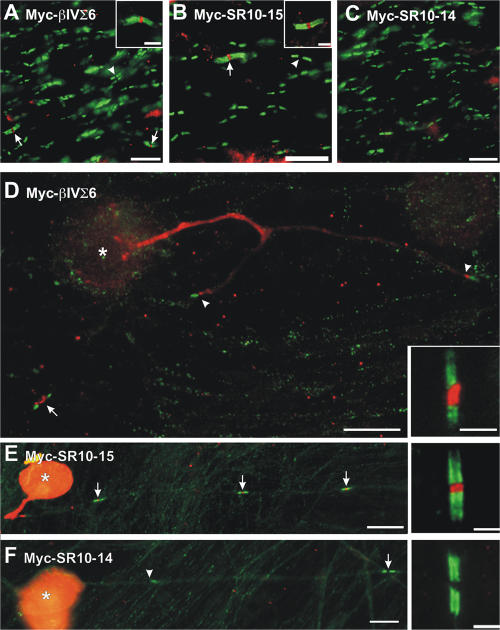
Figure 6.
βIV spectrin localization to nodes of Ranvier in the CNS requires binding to ankG. (A–C) Adenoviruses were injected intravitreally to infect RGCs. Optic nerves were immunostained using anti-Caspr (green) and anti-Myc (red). Myc immunoreactivity was detected at nodes of neurons infected with Myc-βIVΣ6 and Myc-SR10-15 (A and B, arrows and inset), but not Myc-SR10-14 (C). Nodes of Ranvier from uninfected neurons are flanked by Caspr, but do not have Myc immunoreactivity (A and B, arrow). (D–F) Myelinated DRG-Schwann cell cocultures were infected with adenovirus to deliver the βIV spectrin transgenes indicated in each image. 4 d after infection, cocultures were fixed and immunostained using anti-Myc (red) and anti-Caspr (green) to label paranodal junctions and define nodes. Heminodes are indicated by arrowheads, nodes of Ranvier by arrows, and DRG cell bodies by an asterisk. Inset images show higher magnification of a single node of Ranvier from an infected neuron in each culture. Bars: (A–C) 10 μm; (A and B, insets) 3 μm; (D–F) 25 μm; (D–F, insets) 5 μm.

