
Figure 1. Expression of CTIP2 in the mouse fetal skin.
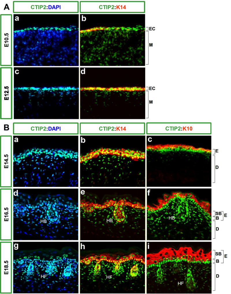
Immunohistochemistry was performed on 10 μm-thick frozen sections of wild type embryos using antibodies directed against CTIP2, K14 and K10. A, CTIP2 (in green) is highly expressed in the ectoderm at E10.5 (upper panel) and E12.5 (lower panel) and is co-localized with the expression of K14 (in red). B, high expression of CTIP2 was observed in the basal cells and upper layers of the epidermis of E14.5 (upper panel), E16.5 (middle panel) and E18.5 embryos (lower panel). K14 and K10 staining (in red) were used to label basal cells and suprabasal layers, respectively. E16.5 and E18.5 stages of development show high expression of CTIP2 in the basal layer of epidermis as well as in the dermis and hair follicles. All sections were counterstained with DAPI (in blue). Images were taken at 40X magnification. Abbreviation: EC- ectoderm, M-mesoderm, E-epidermis, D-dermis, B-basal cell layer, SB- suprabasal cell layers, HB-hair bulb, HF-hair follicle.