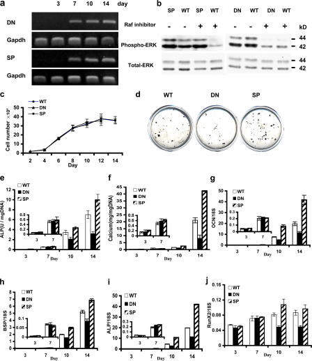
Figure 2.
Altered osteoblast differentiation in calvarial cells from TgMek-dn and -sp mice. Cells were isolated from calvaria of newborn wild-type and transgenic animals. (a) Time course of transgene expression. Cells were plated and grown in differentiating medium for the indicated times before measurement of transgene mRNA by RT-PCR. (b) Mek-dn and -sp transgene expression alters ERK phosphorylation. Cells were grown as in a and harvested after 10 d for measurement of total and phospho-ERK by Western blotting. The indicated groups were treated with the Raf inhibitor ZM336372 2 h before harvest. (c) Transgene expression does not alter cell growth. (d–j) MEK-DN inhibits, whereas MEK-SP stimulates, osteoblast differentiation. The following differentiation markers were measured: mineralized nodules in 14-d cultures (d), alkaline phosphatase activity (e), total cell layer–associated, acid-extractable calcium (f), and OCN, BSP, ALP, and Runx2 mRNA levels (g–j; all measured by real-time RT-PCR). Values are the mean ± the SD of triplicate independent samples.

