Figure 4.
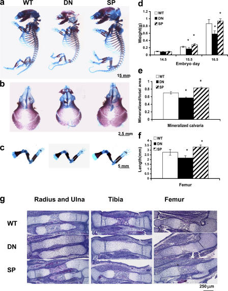
Altered skeletal development in TgMek-dn and -sp mice. (a) Whole mounts of E15.5 skeletons stained with alcian blue and alizarin red. (d) Effects of transgene expression on embryo weights. (b and e) Cranial bones showing differences in mineralization (b) and quantification of mineralized area (expressed as the percentage of total calvarial area; e). (c and f) Hindlimbs showing differences in the size of bones with transgene expression (c) and quantification of femur lengths (f). (g) Histology of long bones from wild-type, TgMek-dn, and -sp mice. Note delay in bony collar and trabecular bone in TgMek-dn embryos. Statistical analysis values are expressed as the mean ± the SD. n = 8 mice/group. *, significantly different from wild type at P < 0.01.

