Figure 1.
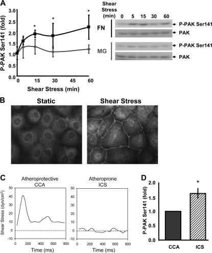
Flow stimulates matrix-specific PAK phosphorylation. (A) BAE cells plated on MG or FN for 4 h were sheared for the indicated times. Phosphorylation of PAK on Ser141 was assessed by immunoblotting total cell lysates with a phosphorylation site–specific antibody. Values are means ± SD normalized for total PAK (n = 3–4). Representative blots are shown. (B) Endothelial cells plated on FN were sheared for 15 min or kept under static conditions, and PAK phospho-Ser141 localization was assessed by immunocytochemistry. Representative images are shown. (C) Shear stress flow profiles for the CCA and the ICS were determined using MRI-generated near-wall velocity gradient profiles of normal carotid arteries. (D) Endothelial cells on FN were stimulated for 24 h with either CCA or ICS flow. Phosphorylation of PAK on Ser141 normalized to total PAK was assessed as described in A. Values are means ± SD after normalization for total protein (n = 3). *, P < 0.05.

