Figure 1.
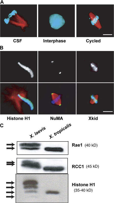
X. tropicalis egg extracts recapitulate major cell cycle events in vitro. (A) CSF-arrested egg extracts prepared from eggs of X. tropicalis and supplemented with X. laevis sperm nuclei and X-rhodamine–labeled tubulin formed “half spindles” in metaphase-arrested extracts, interphase nuclei after release from CSF arrest, and bipolar spindles when induced to reenter metaphase. Microtubules are red and DNA is blue. (B) X. tropicalis extract reactions were stained with fluorescently labeled antibody (α-H1 and α-NuMA), or by immunofluorescence (α-Xkid) using antibodies raised to the X. laevis proteins. Staining patterns recapitulated those in X. laevis reactions. In overlays, the stained protein is green. (C) CSF extracts pooled from eggs of multiple X. laevis or X. tropicalis frogs were blotted for Rae1, RCC1, and histone H1. In X. laevis extracts, all proteins gave multiple bands, presumably because of multiple genes for each protein, whereas X. tropicalis contained single isoforms. Bars, 10 μm.

