
Figure 3.
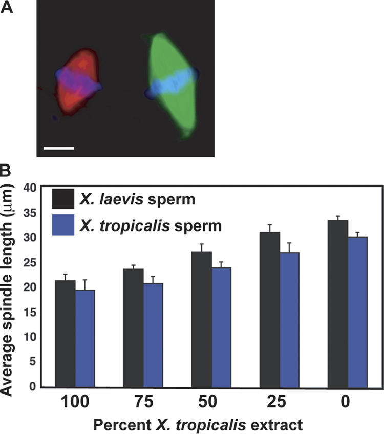
Comparison of spindle length between X. tropicalis and X. laevis. (A) Spindles assembled around X. laevis sperm nuclei in either X. laevis or X. tropicalis egg extracts were visualized using Hoechst dye (blue, DNA) and the incorporation of X-rhodamine tubulin (red microtubules, X. tropicalis), or Alexa Fluor 488 tubulin (green microtubules, X. laevis). Bar, 10 μm. (B) Mixed reactions with the indicated proportion of X. tropicalis extract were combined with X. laevis or X. tropicalis sperm nuclei. Spindle length was measured from pole to pole. A linear relationship was observed between the proportion of X. laevis extract present and spindle length. Error bars are the SD.