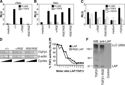
Figure 4.
The RGE mutation does not affect Tgfb1 gene expression, LAP inhibitory function, or latent TGFβ1 processing and secretion. Serum TGFβ1 levels in Tgfb1−/−, Tgfb1+/RGE, and Tgfb1RGE/RGE mice before (A) and after (B) removal of a Neo sequence within the mutated Tgfb1 allele. (C) TGFβ levels in medium conditioned by lung fibroblasts derived from Tgfb1−/−, Tgfb1+/RGE, and Tgfb1RGE/RGE mice. Effects of an anti-TGFβ antibody active against all three TGFβ isoforms (α-TGFβ) and of an antibody that inhibits only TGFβ1 (α-TGFβ1) are shown. Error bars indicate means ± SEM. (D) Tgfb1 gene expression was assessed by semiquantitative RT-PCR using RNA isolated from Tgfb1−/−, Tgfb1+/RGE, and Tgfb1RGE/RGE lung fibroblasts. Reverse-transcribed β-actin mRNA was amplified as a control. (E) Recombinant wild-type and RGE mutant LAP inhibit equally the activity of recombinant TGFβ1, measured with a cell-based luciferase assay. Luciferase activity, expressed as relative light units (RLU), is proportional to TGFβ activity. (F) Wild-type and RGE mutant TGFβ1 cDNAs were expressed in LTBP1S-expressing CHO cells. Secreted proteins were separated by SDS-PAGE in nonreducing conditions and probed with an anti-LAP antibody. Large latent complex (LLC) indicates migration of the LAP–LTBP1 complex.

