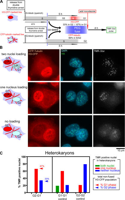
Figure 6.
Passage through mitosis is critical for CENP-A loading in early G1. (A) Schematic of cell synchronization, labeling, and PEG-mediated cell–cell fusion protocol. CENP-A–SNAP cells marked with H3-CFP or CFP-tubulin were sequentially released from a double thymidine block, whereas prior assembled CENP-A–SNAP was quenched. At the time of PEG-mediated fusion H3-CFP and CFP-tubulin cells were in G1 or G2 at the indicated frequencies based on the fraction of cells that had loaded CENP-A–SNAP. After PEG fusion, nocodazole was added to prevent any additional passage through mitosis, and CENP-A–SNAP loading in binucleate heterokaryons was determined after 4 additional hours by TMR-Star labeling. (B) Representative images of binucleate heterokaryons double labeled with H3-CFP and CFP-tubulin, in which both, one, or none of the nuclei has assembled CENP-A–SNAP at centromeres. (C) Frequency of binucleate heterokaryons in which both, one, or none of the nuclei have loaded CENP-A–SNAP at centromeres (TMR-positive nuclei) in fusions of the indicated populations. Arrows in bar graph for G2–G1 cell fusion experiment represent the fraction of H3-CFP cells that were in G1 phase (red) or G2 phase (blue) at the time of fusion. At least 30 binucleate heterokaryons were analyzed in each experiment.

