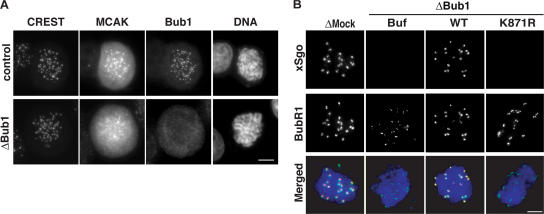
Figure 4.
Bub1 controls localization of MCAK and xSgo to the ICR. (A) Cells treated as in Fig. 2 were stained with antibodies against Bub1, MCAK, and CREST. (B) Chromatin assembled as in Fig. 3 was analyzed by indirect immunofluorescence with antibodies against xSgo (red) and anti-BubR1 (green). DNA was stained as in A (blue).

