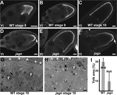
Figure 7.
Transport of Yolkless to the oocyte lateral membrane is reduced in jagn GLCs. (A–F) Wild-type egg chambers (A–C) and jagnQ21X GLCs (D–F) were stained with Yolkless antibodies. Yolkless begins to be transported to the oocyte surface during stage 8 (A). During stages 9 and 10, Yolkless is localized at the oocyte cortex adjacent to follicle cells (B and C). In mutant oocytes (D–F), the intensity of Yolkless staining at the oocyte cortex is reduced. In most egg chambers the distribution of Yolkless is uneven, with the highest level at the posterior region (D and F, arrows). Yolkless enrichment at the posterior region is most evident during stage 9 (D). (G and H) EM images of a wild-type egg chamber (G) and a jagnQ21X GLC (H). Asterisks and y's mark oocyte–follicle cell boundary and yolk granules, respectively. (I) Quantitation of the percentage of yolk area in wild-type egg chambers and jagnQ21X GLCs. Percentage of yolk area was defined as the percentage of area occupied by yolk granules in the ooplasm. N indicates the number of examined oocytes. Bars: (A–F) 20 μm; (G and H) 2 μm.

