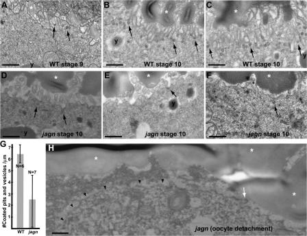
Figure 8.
Endocytosis and cell surface area is reduced in jagn mutant oocytes. EM images of progressive stages of wild-type egg chambers (A–C) and stage 10 jagnQ21X GLCs (D–F). Arrows indicate coated pits and vesicles. y's and asterisks mark yolk granules and vitelline bodies, respectively. In wild-type egg chambers, the oocyte surface has a high density of microvilli, and many coated pits and vesicles are found in the plasma membrane and cortex, especially during stage 10 (A–C). In jagn mutant oocytes, the density of microvilli is reduced to varying degrees, resulting in a decrease of the overall cell surface area (D–F). The number of coated pits and vesicles are also reduced in jagn mutant oocytes (D–F). (G) Quantitation of the number of coated pits and vesicles/micrometer. The number of coated pits and vesicles was counted and divided by the linear length of the examined oocyte surface. N indicates the number of examined oocytes. (H) An EM image of the anterior region of jagnQ21X mutant oocyte. The plasma membrane of the mutant oocyte (arrowheads) is detached from neighboring cells. The number of microvilli (indicated by an arrow) is reduced. Bars, 0.5 μm.

